PHOENIX — Imagine you’ve just been diagnosed with a malignant brain tumor and need surgery. Instead of looking at shadowy black and white 2-D images you can’t decipher, you put on goggles and “walk through” the cancer using augmented reality.
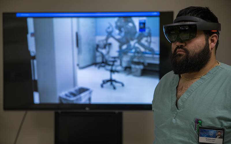
As a result, you can better grasp your disease, said Dr. Matthew Welz, a neurological surgeon at Mayo Clinic Arizona.
“They feel a lot more comfortable going into surgery with this,” Welz said of patients.
The Precision Neurotherapeutics Lab at the clinic in Phoenix is using technology similar to Pokémon Go to ease patient concerns about brain surgery and help doctors find the safest and least invasive route for surgery and treatment.
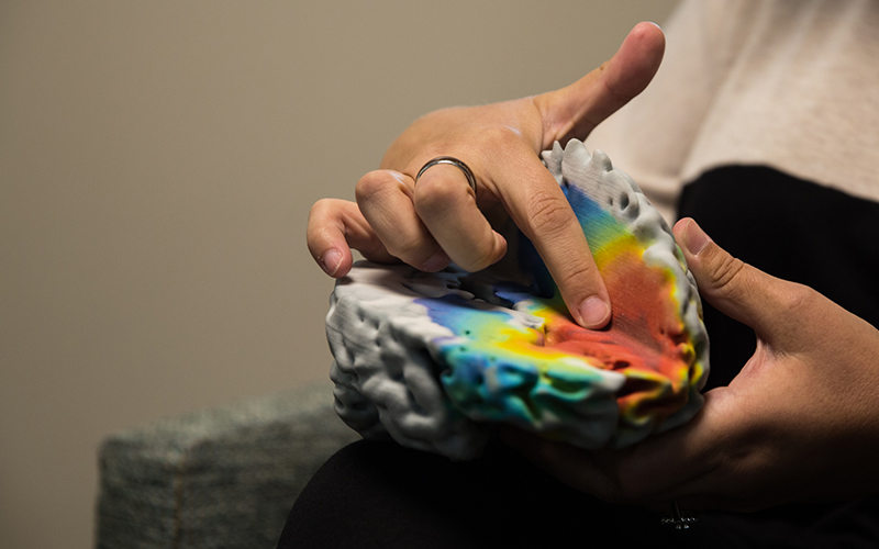
Models from the lab and 3-D simulations create a 360-degree view of the brain, using special goggles, to provide patients with in-depth look at tumors or other abnormalities. It helps them better understand their conditions and possible treatment plans.
Welz said the lab hopes to eventually use the goggles to explain conditions and treatment options to all patients but, for now, limited resources allow only the patients requiring the most critical or complicated procedures to be shown the simulation.
The simulations also predict tumor activity for specific patients, helping health care professionals plan for the best routes of treatment, whether it be radiation, surgery and chemotherapy, said Kristin Swanson, director of Mayo’s Mathematical Neuro-Oncology Lab.
The lab also uses augmented reality to tailor treatment strategies for other conditions, such as scoliosis. 3-D simulations of the spinal column allow surgeons to view overactive curvatures and avoid complications arising from bolts inserted during previous surgeries to straighten the spine.
The simulations help surgeons “do the operation before the operation” to reduce harm while minimizing invasiveness, according to Dr. Bernard Bendok, chair of neurosurgery. He said 3-D imaging displays layers of the brain that 2-D imaging hides, which helps surgeons better differentiate between healthy and cancerous cells.
“What augmented reality is, is the ability through special cameras to superimpose that so that I am actually looking at the real brain. Much like the Pokémon game, I can see additional things layered on top,” Bendok said.
The images, seen through special goggles, show doctors “gradients” of cancerous cells, which allow radiation treatment to target cells in different areas at different intensities, without harming functioning cells.
The lab provides more than just augmented reality. Staffers, led by Swanson, are using high-level math to dramatically change the way doctors are thinking about treatment.

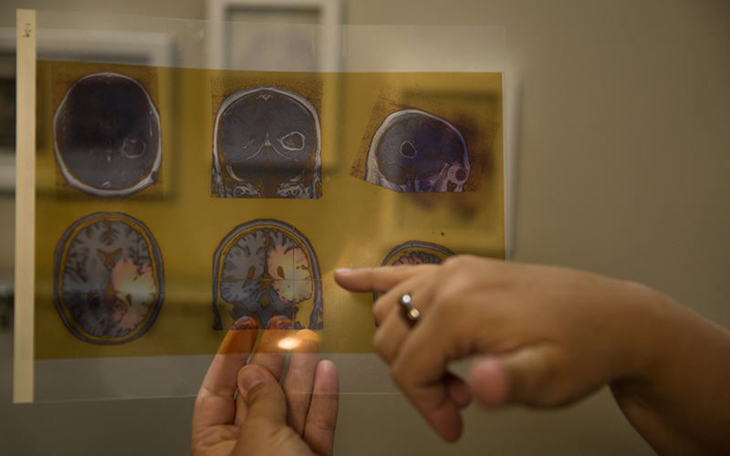
Dr. Kristin Swanson compares images of a brain tumor taken by an MRI (top row) and a heat map created by the Mathematical Neuro-Oncology Lab. “Success is defined by the average right now,” she said, “and we want it not to be defined by the average but instead be defined by the individual patient.” Swanson’s lab predicts brain tumor growth, allowing patients to receive personalized treatment. (Photo by Melina Zuniga/Cronkite News)
Swanson, who earned a doctorate in mathematical biology, started the lab at the clinic in Phoenix about three years ago. She is co-director with Bendok, combining their different specialties: “It’s very personalized medicine meets math.”
She instilled her passion for unique patient care when her father and two brothers died of the same form of cancer. Her father lived eight months, one brother lived 13 months and the other brother died five years after diagnosis.

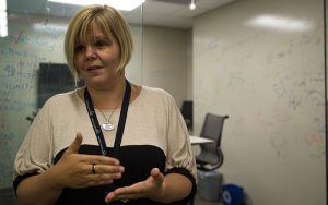
Dr. Kristin Swanson, a mathematical oncologist at Mayo Clinic Arizona in Phoenix, works to predict tumor growth and possible treatment for patients with brain cancer. (Photo by Melina Zuniga/Cronkite News)
“Same diagnosis, similar (tumor) sizes, and similar sets of information,” Swanson said. “They all lived completely differently.”
She said the clinical world uses cohorts like this to find the best average result of treatments to treat the next patient with a similar condition but she wants to evolve health care into personalized treatment.
Personalized maps of the brain not only predict the course of diseases but also keep track of treatment results, Swanson said. Tumor activities, referred to as “hurricanes” in the lab, are tracked as patients undergo treatment, allowing health care professionals to see even the slightest improvements.
“It helps us decide whether we should keep on a path and turn up the volume of that treatment or leave it,” Swanson said.
Bendok agreed with critics who say augmented reality can’t replace doctor-patient relationships or doctors’ “initial-gut instinct” for treatment. But it does complement it.

